Logan Coleman
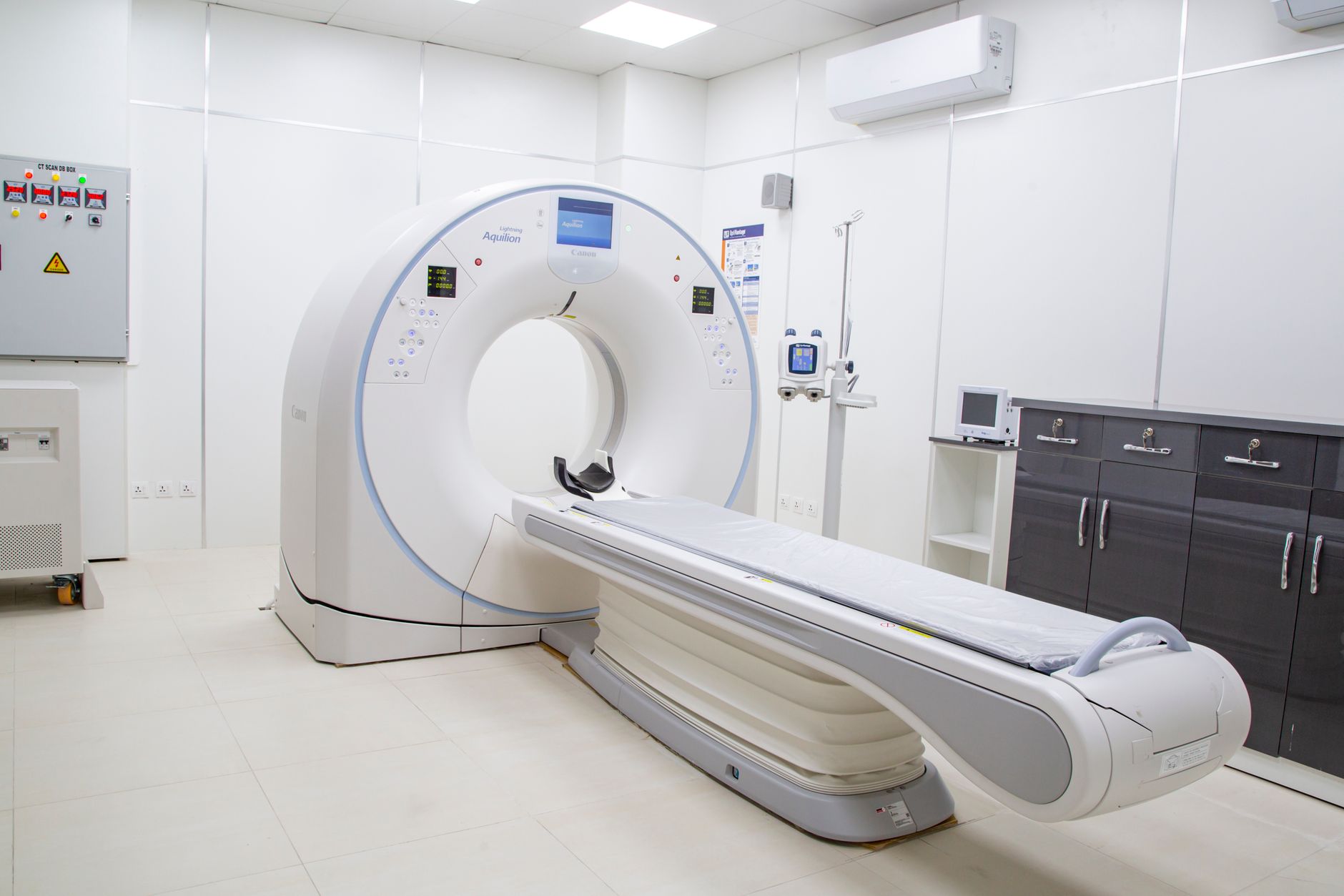
magnetic resonance elastography is making significant strides in oncology, bridging the gap between biomechanics and clinical translation. This innovative technique enhances our understanding of tumor characteristics and treatment responses.
What is Magnetic Resonance Elastography?
Magnetic resonance elastography (MRE) is an advanced imaging technique that measures the mechanical properties of tissues, particularly their stiffness. This non-invasive method utilizes magnetic resonance imaging (MRI) to assess the viscoelastic properties of biological tissues, providing valuable insights in various medical fields, especially oncology.
MRE works by generating shear waves within the tissue, which are then captured by the MRI system. The speed of these waves correlates with the stiffness of the tissue, allowing clinicians to distinguish between healthy and pathological conditions. This is particularly beneficial in oncology, where the characterization of tumors is crucial for diagnosis and treatment planning.
The use of magnetic resonance elastography in oncology offers several advantages:
- Non-invasive: MRE does not require biopsies, reducing patient discomfort.
- Real-time assessment: It provides immediate feedback on tumor stiffness and changes during treatment.
- Enhanced diagnosis: MRE aids in differentiating between tumor types and assessing treatment response.
Applications in Oncology
Magnetic resonance elastography has emerged as a vital tool in the field of oncology, offering significant advantages in the assessment of tumors. This innovative imaging technique enables clinicians to evaluate the mechanical properties of tissues, providing insights that traditional imaging modalities may overlook.
Some key applications of magnetic resonance elastography in oncology include:
- Tumor characterization: By measuring tissue stiffness, magnetic resonance elastography helps differentiate between benign and malignant tumors, facilitating more accurate diagnoses.
- Monitoring treatment response: Changes in tissue elasticity can indicate how well a tumor is responding to therapy, allowing for timely adjustments to treatment plans.
- Staging of cancer: Understanding the biomechanical properties of tumors aids in determining the stage of cancer, which is crucial for planning effective treatments.
With its ability to bridge biomechanics and clinical translation, magnetic resonance elastography is redefining how oncologists approach tumor evaluation and management.
Benefits of Biomechanics in Cancer Treatment
Magnetic resonance elastography (MRE) offers significant advantages in the realm of cancer treatment, particularly by enhancing our understanding of tumor biomechanics. This non-invasive imaging technique provides detailed insights into tissue stiffness, which can be critical for assessing tumor progression and response to therapy.
One of the primary benefits of integrating biomechanics into cancer treatment includes:
- Early Detection: MRE can identify changes in tissue stiffness before conventional imaging methods, enabling earlier intervention.
- Personalized Treatment: By understanding the mechanical properties of tumors, clinicians can tailor treatment plans that are more effective for individual patients.
- Monitoring Treatment Response: MRE allows for real-time assessment of how tumors respond to therapies, offering valuable feedback to optimize ongoing treatment.
- Reduced Need for Biopsies: With accurate biomechanical data, MRE can reduce the reliance on invasive procedures to evaluate tumor behavior.
These compelling benefits illustrate the transformative potential of magnetic resonance elastography in oncology, bridging the gap between biomechanics and clinical outcomes.
Future of Magnetic Resonance Techniques
The future of magnetic resonance techniques, particularly magnetic resonance elastography, holds significant promise for advancing oncology. As research continues to unfold, the integration of these innovative imaging methods into clinical practice is expected to enhance diagnostic accuracy and treatment planning.
One of the most exciting prospects is the potential for real-time monitoring of tumor responses to therapy. This capability could lead to more personalized treatment approaches, allowing oncologists to adjust therapies based on the biomechanical properties of tumors as they change over time.
Furthermore, ongoing studies are exploring the application of magnetic resonance elastography beyond traditional oncology, possibly extending its benefits to other medical fields. The evolution of these techniques may also pave the way for improved patient outcomes, as they offer less invasive options for assessing tumor characteristics.
As we look ahead, continued collaboration between researchers and clinicians will be essential in translating these advancements into routine clinical use, ensuring that the full potential of magnetic resonance elastography is realized in the fight against cancer.
Clinical Case Studies and Results
Recent clinical case studies have highlighted the effectiveness of magnetic resonance elastography in the assessment and treatment of various cancers. By providing detailed insights into tissue stiffness, this advanced imaging technique has proven to be invaluable in characterizing tumors and monitoring treatment response.
In one notable study involving patients with liver cancer, magnetic resonance elastography facilitated the early detection of significant changes in tumor stiffness, which correlated with treatment efficacy. This early detection allowed for timely adjustments in therapy, ultimately improving patient outcomes.
Another case demonstrated its utility in breast cancer management, where magnetic resonance elastography helped differentiate between benign and malignant lesions, thereby reducing unnecessary biopsies. The ability to assess tumor biomechanics also aids clinicians in tailoring personalized treatment plans based on individual tumor characteristics.
These clinical findings underscore the potential of magnetic resonance elastography to bridge the gap between biomechanics and clinical translation, enhancing the precision of cancer care and paving the way for future research in oncology.
Photo by contact me +923323219715 on Pexels